
Why predict what we can now measure?
10 September 2026 / 12:36 pm
What the biggest imaging study of silent atherosclerosis tells us about the tools we use to prevent heart attacks
In August the New England Journal of Medicine published the baseline results of REACT, a Danish-Spanish cohort of 16,808 adults aged 18 to 70 with no known cardiovascular disease (1). Every participant underwent three-dimensional ultrasound of the carotid and femoral arteries and CT angiography of the coronary arteries.
It is the most complete atlas of silent atherosclerosis across adult life we have ever had.
The headline is that 57% of these apparently healthy people already had plaque somewhere. But another number matters even more. When the investigators applied SCORE2, the risk calculator that European guidelines use to decide who gets treated, only 1.9% of participants with subclinical atherosclerosis were classified as high risk. Nineteen in twenty were not.
That is not because SCORE2 is a bad risk score. It was designed to predict cardiovascular events, not to diagnose atherosclerosis. But it exposes a widening gap between estimating who might have an event over the next decade and identifying who already has the disease we are trying to prevent.
The disease is everywhere
REACT stratified enrolment by age and sex, giving an unusually clear picture of how atherosclerosis develops through adult life.
Roughly one in thirteen people in their twenties already had plaque. By their thirties it was a third of men and a fifth of women. By their sixties nine in ten had plaque somewhere, and only 1.9% of men aged 60 to 70 had completely clean arteries.
Women started later, but prevalence rose steeply after 40, in a curve that overlaps strikingly with the menopausal transition.
Plaque burden increased roughly exponentially with age in every vascular territory. What began as a single carotid or femoral lesion in a thirty-year-old became multi-territory disease in most sixty-year-olds. Among men in the oldest group, 56% had plaque in all three vascular beds.
Atherosclerosis, then, is not something most of us are at risk of developing. By later life, most of us already have it. The questions that matter are how much disease, where, and how active is it.
What a risk score actually does
SCORE2, QRISK, PREVENT and their predecessors are regression models derived from populations. They combine age, blood pressure, cholesterol and smoking to estimate the probability that someone with a similar profile will have a cardiovascular event over a defined period. They estimate an outcome, but they do not measure the disease.
Age carries enormous weight in every risk model. So a younger person can have decades of exposure to atherogenic risk factors, and established plaque, while retaining a reassuringly low ten-year predicted risk. An older person can cross a treatment threshold on age alone with very little atherosclerosis to show for it.
The REACT study makes the problem visible. SCORE2 discriminated only modestly between people with and without plaque, and it is not calibrated at all below 40, exactly where the gap between short-term risk and early disease is widest.
We are estimating something we can now look at.
Measure, don’t just estimate
The alternative is not a better prediction model. It is measurement. Imaging and biomarkers tell us whether disease is present in an individual, how extensive it is and, increasingly, how active it is.
REACT offers three additional practical lessons. First, isolated coronary disease was uncommon, found in at most 9% of men and 5% of women. Among people with coronary plaque, 82% also had plaque in the carotid or femoral arteries, and carotid ultrasound gave the highest diagnostic yield of any single site.
Second, a calcium score of zero is not the absence of coronary atherosclerosis, particularly in younger adults. Among 30-to-39-year-olds with coronary plaque, 42% of men and 48% of women had a calcium score of zero. Their plaque had simply not calcified yet. In Jersey we have stopped measuring calcium score in isolation for risk assessment, yet calcium scoring has just been strongly recommended in recent North American guidelines. CAC=0 remains a powerful prognostic marker but if the question is whether a person has coronary plaque, calcium scoring cannot answer it.
Third, finding plaque is not enough. If nine in ten sixty-year-olds have some atherosclerosis, “plaque present” discriminates very little. The value of imaging lies in grading disease including plaque burden, the number of territories involved, plaque composition and biological activity.
Seeing inflammation
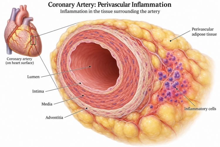
This is where coronary inflammation provides additional vital information. Atherosclerosis is an inflammatory disease. Plaque does not rupture because it is large; it ruptures because it is inflamed. Yet until recently we had no way of seeing inflammation in an individual artery.
The measurement of fat attenuation index (FAI) has changed that. It rests on a simple biological observation. The fat that wraps the coronary arteries is not inert packing; it is in constant conversation with the vessel wall. When the wall becomes inflamed, it releases signals that stop the adjacent fat cells from storing lipid. The fat becomes smaller, more watery, and on a standard CT angiogram this shows up as a measurable shift in attenuation. The inflamed artery, in effect, leaves a fingerprint on the tissue around it. No extra scan, no extra contrast, no extra radiation.
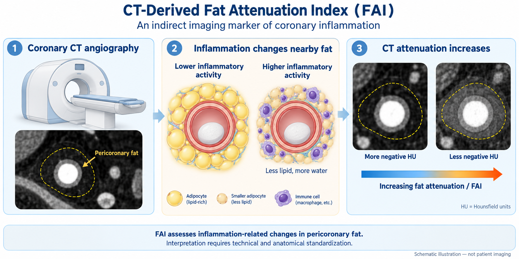
The evidence base for the use of FAI is now substantial. The first large validation in The Lancet in 2018 showed that a high perivascular fat attenuation predicted cardiac death several-fold over and above calcium score and the degree of stenosis (2). ORFAN, published in the same journal in 2024, followed just over 40,000 patients from eight NHS hospitals for a median of 2.7 years (3). Four in five had no obstructive coronary disease, and yet that group accounted for nearly twice as many cardiac deaths and major events in absolute terms as those with obstructive disease. Inflammation, measured from the same CT the patient was having anyway, identified who among the non-obstructive majority was at risk, independent of the usual risk factors and of plaque burden itself.
ORFAN also went a step further than a single biomarker. The inflammation signal was combined with plaque characteristics and conventional risk factors into an AI-derived estimate of absolute risk over the following years. That distinction matters. A stenosis grade tells you what the artery looks like; a risk of 15% over eight years tells you what to do about it.
That finding matters for REACT’s population. The people REACT describes are exactly this non-obstructive majority: plaque present, arteries open, risk score reassuring. The question for them is not whether they have disease but whether it is quiet or active, and inflammation is the first non-invasive measure that speaks directly to that.
It also changes what clinicians do. In Jersey we took 79 consecutive patients whose CT angiograms were entirely clean, with no plaque at all, and ran the inflammation analysis on the same scans (4). Compared with QRISK3, the inflammation-based risk score reclassified nearly half of them. A third had their management changed as a result, almost all by starting a statin they would otherwise not have been offered. These were people who had just been told their arteries looked normal. The picture said no disease; the biology said otherwise.
There are some caveats. The measurement currently needs a contrast CT angiogram, although emerging data from the Oxford group suggests that might not always be the case, so it belongs at the top of a prevention pathway rather than the bottom. Standardisation across scanners and vendors is still maturing and no randomised trial has yet shown that treating on the basis of an inflammation score changes outcomes. But as a way of separating the plaque that will sit harmlessly for decades from the plaque that will not, it is the most promising tool we have. The future therefore lies not in detecting plaque but in separating quiet disease from active disease.
A pathway
Put these observations together and a tiered prevention pathway emerges, one that a small health system such as Jersey could deliver end to end and starting with blood tests. Lipoprotein(a) is largely genetically determined and needs measuring once. ApoB directly counts the atherogenic particles in circulation. Both identify risk that conventional calculators and lipid measurements miss.
Then imaging. A carotid and femoral ultrasound at age 40, earlier for those with a strong family history, a high Lp(a) or other major risk factors, establishes whether atherosclerosis is already present. Coronary CT angiography is reserved for those in whom the detail would change management or for older patients, quantifying plaque burden and composition. Coronary inflammation with FAI adds a further layer of stratification for those with coronary disease.
Treatment intensity then follows measured disease rather than a risk threshold. For many that means lifestyle change and a statin. For those with extensive or biologically active disease, it means more including intensive lipid lowering, tighter blood-pressure control, metabolic therapy or anti-inflammatory treatment as the evidence for each matures.
There is one further advantage that no risk score can offer. A patient who has seen their own arteries is more likely to take their tablets. Adherence to some medicines can be poor but adherence after a picture of your own plaque is a different matter.
What we do not yet know
REACT is cross-sectional and describes prevalence, not events. The evidence that subclinical plaque predicts cardiovascular outcomes comes from other cohorts, including BioImage, MESA, SCAPIS and Copenhagen, where imaging-defined disease consistently adds prognostic information beyond conventional risk factors.
We still however lack a large completed randomised trial showing that systematic imaging of asymptomatic adults, followed by imaging-guided treatment, reduces myocardial infarction or cardiovascular death. Until those trials report, the case for moving from estimation to measurement is compelling rather than proven. REACT nonetheless forces an uncomfortable question. Our prevention systems are very good at estimating short-term event risk. They were never designed to tell us whether atherosclerosis is present. For most of medical history that distinction hardly mattered, because we had no practical way of seeing the disease. We do now.
The question is not whether we can predict cardiac risk better. It is why we are still predicting something we can measure.
- Bundgaard H, García-Lunar I, Kofoed KF, et al. REACT Investigators. Prevalence of Silent Atherosclerosis across Adult Life. N Engl J Med 2026 doi: 10.1056/NEJMoa2609059
- Oikonomou E, Marwan M, Desai M et al.Non-invasive detection of coronary inflammation using computed tomography and prediction of residual cardiovascular risk (the CRISP CT study): a post-hoc analysis of prospective outcome data. The Lancet 2018;392:929-939
- Chan K, Wahome E, Tsiachristas A et al. Inflammatory risk and cardiovascular events in patients without obstructive coronary artery disease: the ORFAN multicentre, longitudinal cohort study. The Lancet 2024;403: 2606-2618
- Henry JA, Black SM, Mitchell OG, et al. Coronary inflammation and AI-Risk scores from cardiovascular computed tomography: impact on risk prediction and clinical management in a real-world setting. European Heart Journal – Imaging Methods and Practice 2024;2(24): qyaf031
More Posts
19 November 2025
Trust and Hope: Building Jersey’s New Era of Health Care
7 May 2025
A digital health diary revisited
16 March 2024
Developing a new strategy for healthy Islanders
4 February 2024
A digital health diary for Jersey
27 December 2022
Utilising artificial intelligence in cardiology practice
12 June 2022
Under the bonnet – time for your heart’s MOT
12 March 2022
What is heart valve disease and what does that mean for me?
12 January 2022
Preventing heart disease using augmented digital health intelligence
1 October 2021
How heart health has been impacted by COVID-19
1 August 2021
The importance of the rhythm of your heart
21 June 2020
What is atrial fibrillation?
8 April 2020
Coronary heart disease and risk of heart attacks
11 January 2020
The Era of Immersive Health Technology
22 September 2019
The heart and stress
18 August 2019
Screening for Atrial Fibrillation and the Role of Digital Health Technologies