Cardiac magnetic resonance imaging (MRI)
23 May 2021 / 3:23 pm
What is it ?
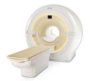
A cardiac MRI is an investigation to image your heart structure and function in detail using a strong magnetic field. A cardiac MRI is particularly useful to help with the diagnosis of certain heart muscle abnormalities such as cardiomyopathies.
How is it performed ?
You will be asked to lie on a special couch so that the MRI can scan around your chest. The machine can be noisy so you will be given headphones to wear. You will need to hold your breath for 10 seconds to keep your chest still. Sometimes an injection of a contrast (gadolinium) is given to highlight parts of the heart.
How long does it take ?
Up to an hour.
Are there any risks ?
There is no radiation risk with an MRI. You need to tell the MRI department if you have any implants or metal objects in your body, particularly a pacemaker. Allergic reactions to the contrast are uncommon.
What happens next ?
After you have had your cardiac MRI, the images will be analysed and a report generated, usually within seven days.