Myocardial perfusion scan
23 May 2021 / 4:27 pm
What is it ?
A myocardial perfusion scan is a test to determine if there is any restriction in blood supply to the heart muscle due to coronary heart disease. A normal result means that the risk of a heart attack over the next few years is extremely small.
How is it performed ?
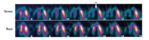
The heart is stressed, either by getting you to exercise gently or by the use of a drug into your vein. When your heart rate has increased sufficiently, or the drug infusion is complete, a radioactive tracer is injected into your blood stream. This is fixed in your heart. You will then be positioned in a scanner to determine if parts of your heart muscle are not getting adequate blood supply.
How long does it take ?
The initial test takes about thirty minutes. If there is any abnormality detected in the first scan, then you may be asked to return for a rest scan a few hours later so that a comparison can be made.
Are there any risks ?
The amount of radiation used with this test is extremely small and the test would only be arranged if it was felt that the benefit from the scan outweighed any risk to your health. If you are taking the drugs dipyridamole, aminophylline or theophylline then they should be stopped at least 24 hours before the test.
What happens next ?
Once you have had your test, the images will be analysed and a report generated. This usually takes about a week.