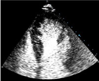
Contrast echocardiogram
23 May 2021 / 4:25 pm
What is it ?
A contrast echocardiogram is an ultrasound investigation of the heart. Sound waves are reflected of structures in the heart generating a 2D and 3D picture of the heart. A contrast agent (such as Sonovue® or agitated saline / salty water) is injected into the blood stream to make the pictures clearer. A contrast echocardiogram test is particularly helpful for examining the heart muscle in detail and for identifying any holes in the heart.

How is it performed ?
The test is performed in a private room as you will need to take off the clothes from your upper body. You are welcome to bring along a friend or relative who can sit in with you. You will need to lie on a couch and a small probe will be placed on your chest with lubricating jelly to improve the picture quality. You may be asked to roll over or lie flat and to hold your breath at times. A small plastic tube is inserted into one of your veins. The contrast is injected into this to improve image quality.
How long does it take ?
A detailed contrast echocardiogram usually takes up to hour to acquire. The report can then take up to an hour to generate.
Are there any risks ?
The ultrasound is harmless. The contrast agent is rapidly dispersed and allergic reactions are uncommon.
What happens next ?
Once you have your contrast echocardiogram, the images will be analysed and a report generated, usually within seven days.